

Retinal Tear Causes – How They Can Be Managed?
The retina is the light-sensing tissue lining the inside of the eye. Made up of ten layers of different types of cells, the retina is crucial for vision. Any damage to this structure from trauma or disease, including a severe retinal tear, can cause permanent loss of vision in that particular part of the eye.
What is a Retinal Tear?
A retinal tear is exactly as it sounds – a rip or tear in the fragile tissue of the retina. This is different from a retinal detachment, where a section of the retina peels away entirely from the underlying structures of the eye, however, it is not impossible that a retinal tear causes a detachment, which must then be treated urgently with retinal surgery.
The retina contains no pain nerves and so a retinal tear causes no physical discomfort or pain. Some instances of a retinal tear may in fact present with no symptoms at all, particularly if the tear is small or very far out in the periphery of the eye where we are less aware of changes to our vision. In other cases, a retinal tear causes symptoms including:
- A sudden appearance of floaters in the vision of the affected eye – these can be described as little black dots or spots, or cobwebs. Some people in fact think they’re seeing a swarm of tiny black insects before realising the floating specks are inside their own eye.
- Flashing lights in the vision – the medical term for this is photopsia, and often appears as a lightning arc out of the corner of your eye. The flashes may be recurrent and persist for some time.
- Hazy vision – if the tear has disrupted some retinal capillaries, the resultant bleed into the space of the eyeball can cause a blurry patch in your vision.
If the retinal tear causes a detachment you may notice more dramatic symptoms, such as the sensation of a dark shadow or curtain falling across part of your vision and obscuring that part of your sight. If you experience this it is important to promptly seek the attention of an eye specialist experienced in retinal surgery.
Retinal Tear Causes
Most instances of a retinal tear are idiopathic, meaning they occur spontaneously with no identifiable underlying reason. There are certain factors which may put you at a higher risk of developing a retinal tear, including: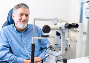
- Older age
- A family history or retinal tears or detachments
- A history of trauma to the eye or head
- Myopia, also known as near-sightedness or short-sightedness, as this results in a thin retina
- Lattice degeneration, which involves thinning of areas in the peripheral retina
- A history of an eye operation, including cataract surgery
One of the common retinal tear causes can be attributed to a normal age-related change called a posterior vitreous detachment (PVD). The vitreous humour is a clear jelly-like substance that fills the back chamber of the eyeball, and is anchored to certain points along the retina.
As we age, the vitreous begins to liquefy, resulting in its solid molecular structure slowly collapsing. While this process is entirely normal, occasionally the adhesion between the vitreous gel and the retina is too tight and so does not separate easily as the vitreous liquefies. The tension on this anchor point may be enough to tug so hard on the retina that it causes a tear in the tissue, or possibly even a detachment.
An eyecare practitioner such as an optometrist or ophthalmologist will often find a retinal tear incidentally during a routine eye exam. This can be achieved by using dilating eye drops to widen the pupil and utilising an illumination and magnification system to view the retina, such as a slit lamp biomicroscope or a binocular indirect ophthalmoscope. If there is a haemorrhage obscuring part of the retina where a tear or detachment is suspected, an imaging device such as an ultrasound may be required to view the retina behind the bleed.
Retinal Surgery
The main determining factor when it comes to deciding whether retinal surgery is warranted for a tear is its potential to deteriorate into a vision-threatening retinal detachment. Retinal tears that are small, peripheral, and oriented in a certain way may be deemed low-risk, and therefore can be monitored carefully without intervention. Some tears in fact self-heal by naturally forming their own scarring, thereby resealing themselves.
If a tear is thought to have significant potential to lead to a retinal detachment, also when considering a person’s other risk factors such as the presence of high short-sightedness, the eye specialist may recommend either a laser procedure called photocoagulation or a freezing technique known as cryopexy in order to seal the edges of the tear to prevent it from deteriorating.
In a way, both these treatments function like spot-welding, creating microscopic patches of controlled scarring that re-stick the torn retina to the underlying tissue. Photocoagulation is done under a topical anaesthetic in the ophthalmologist’s office and only takes several minutes. There is very little recovery time required afterward, if any at all, and many people resume their usual activities immediately. In the case of cryopexy, there may be a period of recovery over a couple of weeks.
It’s important to understand that successful treatment for a retinal tear doesn’t preclude you from experiencing another tear, either in the same or opposite eye. Your eye specialist will recommend you maintain regular and frequent eye exams with your eyecare practitioner. At Mornington Peninsula Eye Clinic, our ophthalmologists examine the retina in detail and explain the treatment options for a retinal tear before you decide. To arrange an appointment, please call us on (03) 9070 3580.
Note: Any surgical or invasive procedure carries risks. Before proceeding, you should seek a second opinion from an appropriately qualified health practitioner.


Leave a Reply
Want to join the discussion?Feel free to contribute!