
Understanding The Pathophysiology Of Glaucoma: What It Is, Risk Factors, Types, And Treatments
Glaucoma is a compilation of eye diseases that damage the optic nerve, the crucial pathway delivering ocular information from the eye to the brain. This damage, often caused by intraocular pressure elevation, leads to gradual vision loss and may eventually lead to irreversible blindness if left untreated. This article delves into the pathophysiology of glaucoma, explores the risk factors and different types, and discusses treatment options and how to choose the right ophthalmologist for your needs.
The Delicate Balance: Pathophysiology Of Glaucoma
The healthy eye maintains a precisely regulated internal pressure, termed intraocular pressure (IOP), which is critical for preserving its spherical form and ensuring optimal function of its delicate internal structures. Aqueous humour, a transparent, nourishing fluid, is continuously produced by the ciliary body, a structure situated posterior to the iris.
 This fluid fills the anterior chamber, the space between the iris and cornea at the front of the eye. Aqueous humour constantly circulates within the anterior chamber, bathing and providing essential nutrients to the eye’s internal components.
Maintaining a meticulous balance of aqueous humour is paramount for healthy vision. This equilibrium is achieved through a well-orchestrated interplay between production and drainage. The ciliary body acts as a continuous source of aqueous humour, and this fluid subsequently exits the eye via a specialised drainage network known as the trabecular meshwork.
Located at the anterior chamber angle, where the iris meets the cornea, the trabecular meshwork functions akin to a sieve, facilitating the outflow of aqueous humour from the eye into collector channels that ultimately drain into the bloodstream. In a healthy eye, production and drainage of aqueous humour are meticulously balanced, resulting in a normal IOP typically ranging from 11 to 21 mmHg.
Glaucoma disrupts this delicate homeostasis. The most prevalent cause of glaucoma is elevated IOP, which can arise from two primary mechanisms:
- Impeded Drainage: The trabecular meshwork can become obstructed or dysfunctional, hindering the outflow of aqueous humour. This obstruction leads to a progressive accumulation of pressure within the eye.
- Increased Production: Excessive production of aqueous humour by the ciliary body can also contribute to high IOP. This phenomenon can occur due to various underlying conditions or even as a primary abnormality.
Elevated IOP poses a significant threat to the optic nerve, the crucial pathway transmitting visual information from the retina to the brain. The optic nerve head, the area from which the optic nerve leaves the posterior aspect of the eye, is particularly susceptible to pressure-induced damage. Chronic high IOP exerts undue stress on the delicate retinal ganglion cells (RGCs) residing within the optic nerve head.
These RGCs are specialised neurones responsible for converting light signals into electrical impulses that move along their axons (nerve fibres) to the brain. Sustained compression from an elevated IOP can lead to retinal ganglion cell axons’ damage and retinal ganglion cell death.
This progressive loss of RGCs and their axonal connections ultimately results in characteristic alterations in the optic nerve head appearance and, more importantly, irreversible vision loss.
A comprehensive understanding of the intricate mechanisms underlying the pathophysiology of glaucoma is fundamental for the development of effective treatment strategies.
Who’s More Likely To Develop Glaucoma? Risk Factors To Recognise
Glaucoma isn’t a single disease but a spectrum of conditions marked by progressive optic nerve damage and vision loss. Several established risk factors significantly increase susceptibility. Understanding these is crucial for early detection and intervention.
- Elevated Intraocular Pressure (IOP): While not everyone with high IOP develops glaucoma, it’s a cause for concern. Chronically high IOP stresses the optic nerve, contributing to vision loss.
- Age: The risk of glaucoma demonstrably increases with age, particularly after 60.
- Family History: Having a close relative with glaucoma significantly increases your own risk.
- Ethnicity: Individuals of African descent are more likely to develop glaucoma, particularly primary open-angle glaucoma.
- Medical Conditions: Certain diseases, like diabetes and high blood pressure, can elevate glaucoma risk.
- Thin Corneas: Individuals with thinner corneas may be more susceptible to optic nerve damage at lower IOP levels.
- Previous Eye Injuries: Significant eye injuries can damage drainage structures or increase IOP, raising glaucoma risk.
By recognising these risk factors and undergoing regular eye exams, you can take proactive steps towards protecting your vision and ensuring early detection of glaucoma.
Unveiling The Different Types Of Glaucoma
Glaucoma encompasses a spectrum of eye diseases characterised by progressive optic nerve damage and vision loss. Glaucoma manifests in various forms, each with distinct characteristics and presentations. Here, we delve into the four major types of glaucoma:
Primary Open-Angle Glaucoma (POAG)
This most common type develops gradually with minimal initial symptoms. The trabecular meshwork becomes progressively less effective in draining aqueous humour, leading to slow-onset vision loss, typically affecting peripheral vision first.
Angle-Closure Glaucoma (ACG)
This type arises due to a sudden blockage of the anterior chamber angle, often by the iris. This blockage dramatically increases IOP, causing severe eye pain, redness, blurred vision, and sudden vision loss. It requires immediate medical care to prevent permanent vision loss.
Normal-Tension Glaucoma
In this form, optic nerve damage and vision loss occur despite IOP measurements within the normal range. It’s believed that these individuals may be more susceptible to nerve damage at lower pressures.
Secondary Glaucoma
This type of glaucoma arises due to underlying medical conditions like uveitis (eye inflammation) or trauma that damage the drainage structures or increase IOP.
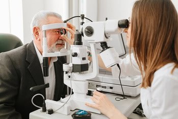
Unveiling The Silent Thief: How Glaucoma Is Diagnosed
Glaucoma, a stealthy thief of sight, often progresses without noticeable symptoms in its early stages. Early detection and intervention are paramount for preserving vision. This section delves into the comprehensive diagnostic process employed by ophthalmologists to identify and classify glaucoma.
Detailed Medical History
The initial consultation involves a detailed discussion of your medical history, including any family history of glaucoma, previous eye conditions or injuries, and current medications.
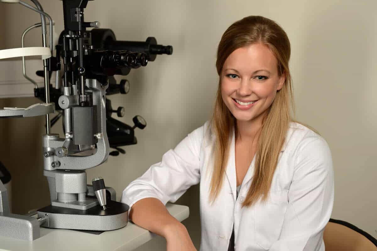
Ocular Examination
A comprehensive eye exam is essential for evaluating potential signs of glaucoma. This examination typically includes:
- Visual Acuity Testing:
 This standard test measures your ability to see at various distances, helping to identify any vision loss associated with glaucoma.
 - Slit-Lamp Examination: Using a specialised microscope with a high-intensity light source, the ophthalmologist examines the structures of your eye in detail, including the cornea, iris, lens, and optic nerve. This examination can reveal signs of damage or abnormalities suggestive of glaucoma.
- Tonometry: This test measures intraocular pressure (IOP), a crucial indicator of glaucoma risk. Several methods are available, including applanation tonometry (a gentle puff of air on the cornea) and rebound tonometry (a quick, simple touch to the cornea).
- Ophthalmoscopy: With dilated pupils, the ophthalmologist examines the back of your eye, focusing on the optic nerve head. The appearance of the optic nerve, including its colour, cupping (excavation), and blood vessel patterns, provides valuable clues about the potential damage caused by glaucoma.
- Gonioscopy: In some cases, this specialised examination may be necessary to assess the drainage angle (the angle formed by the iris and cornea) where aqueous humour exits the eye. This evaluation helps determine if the angle is open or closed, influencing the type of glaucoma.
 This standard test measures your ability to see at various distances, helping to identify any vision loss associated with glaucoma.
Visual Field Testing
This test assesses peripheral (side) vision and helps identify potential blind spots or areas of vision loss that may occur in glaucoma. Various techniques are available, including automated perimetry and confrontational visual field testing.
Imaging Tests
In some situations, your ophthalmologist may employ advanced optical coherence tomography (OCT) for a more detailed evaluation. This non-invasive imaging technology provides high-resolution cross-sectional images of the optic nerve head, allowing for precise nerve fibre layer thickness measurement. This information is valuable in detecting and monitoring glaucoma progression.
Addressing The Root Cause: How To Deal With Glaucoma’s Pathophysiology
Glaucoma, while not curable in the sense of reversing existing nerve damage, is a highly treatable condition. Our primary therapeutic goal is to address the underlying pathophysiology—an elevated intraocular pressure (IOP)—and prevent further optic nerve degeneration. Here’s an in-depth exploration of the treatment modalities employed:
Medications
Eye drops are the cornerstone of initial treatment for most glaucoma patients. They offer a simple and non-invasive approach to managing IOP. Several classes of medications work by targeting different aspects of aqueous humour dynamics:
- Prostaglandin Analogues: These medications act on the outflow pathway, promoting increased drainage of aqueous humour through a network of channels outside the trabecular meshwork. They are typically well-tolerated and offer a long-lasting IOP reduction.
- Beta-Blockers: These medications decrease the production of aqueous humour by the ciliary body. They are particularly beneficial for individuals with conditions like ocular hypertension, where beta-blockers may already be part of their overall medical regimen.
- Carbonic Anhydrase Inhibitors: These medications act systemically to decrease aqueous humour production. They are typically used as an adjunct to other topical medications or in specific situations where they are not tolerated or effective.
The choice of medication and dosage are personalised based on the type and severity of glaucoma, IOP level, potential side effects, and individual patient factors. In some cases, a combination of different medication classes may be necessary to achieve optimal IOP control.
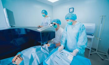
Laser Trabeculoplasty
This minimally invasive outpatient procedure utilises a laser to target the trabecular meshwork. The laser creates microscopic burns within the meshwork, improving its drainage capacity and facilitating increased aqueous humour outflow. This procedure is often used for primary open-angle glaucoma (POAG) patients who are not adequately controlled with medications.
Minimally Invasive Glaucoma Surgery (MIGS)
This is a growing category of surgical procedures designed to offer a less invasive approach compared to traditional glaucoma surgeries. MIGS procedures utilise microscopic implants or devices to improve aqueous humour outflow through various mechanisms. They are often performed in conjunction with cataract surgery or as a standalone procedure for mild to moderate glaucoma. Some advantages of MIGS include:
- Shorter operating time
- Reduced risk of complications
- Faster recovery time
Incisional Surgery
When medications and laser treatment fail to adequately control IOP, surgery becomes necessary. There are two main types of glaucoma surgeries:
- Trabeculectomy: This traditional surgical approach creates a new drainage channel for aqueous humour in the sclera (the white part of the eye). This channel bypasses the obstructed trabecular meshwork and allows fluid to drain from the eye, reducing IOP.
- Drainage implants: These are tiny tubes implanted into the eye that create a new drainage channel for aqueous humour. This approach is gaining popularity due to potentially less invasive techniques and a shorter surgical duration than trabeculectomy.
Know The Signs: How To Recognise Glaucoma
Glaucoma is oftentimes called the “silent thief of sight” because the early stages often progress without noticeable symptoms. However, some signs may indicate the development of glaucoma, especially in later stages:
- Peripheral Vision Loss: This is a common early sign, with gradual narrowing of your side (peripheral) vision. You might miss objects on your side or feel the need to turn your head further to see things.
- Tunnel Vision: In advanced glaucoma, central vision may also deteriorate, leaving only a narrow central field of view, like looking through a tunnel.
- Sudden Vision Loss: This is more common in angle-closure glaucoma, where a blockage in the anterior chamber angle rapidly increases IOP.
- Haloes Around Lights: You might see rainbow-coloured rings or halos around lights, especially at night.
Early detection is crucial for preserving vision. Regular eye examinations, especially for higher-risk patients, are essential for identifying glaucoma before significant damage occurs.
Choosing The Right Ophthalmologist For Your Glaucoma Treatment
Selecting the right ophthalmologist for your glaucoma care is vital. Look for an ophthalmologist specialising in glaucoma management. Here are some factors to consider:
- Experience: Choose an ophthalmologist with extensive experience diagnosing and treating glaucoma, particularly your specific type.
- Technology: Ensure the ophthalmologist’s practice has access to advanced diagnostic tools for glaucoma evaluation.
- Treatment Options: Enquire about the range of treatment options offered, including medications, laser procedures, and surgical expertise.
- Communication style: Choose an ophthalmologist who clearly explains your exact condition, treatment options, and potential risks and benefits.
 Choose an ophthalmologist with extensive experience diagnosing and treating glaucoma, particularly your specific type.
A good doctor-patient relationship is key for successful glaucoma management. Ask questions and sound off your concerns to ensure you feel confident in your ophthalmologist’s care.
Frequently Asked Questions
Can I go blind from glaucoma?
If left untreated, glaucoma can be the leading cause of irreversible blindness. Early detection and consistent treatment can significantly reduce the risk of vision loss.
What happens if I stop taking my glaucoma medication?
Glaucoma medications are crucial for lowering IOP and preventing further optic nerve damage. Stopping your medication can cause IOP to rise, accelerating disease progression and increasing your likelihood of experiencing vision loss.
I have high blood pressure. Does this increase my risk of glaucoma?
Yes, high blood pressure is a risk factor for glaucoma. Maintaining good blood pressure control is crucial to reducing overall risk.
I’m worried about glaucoma, but I don’t have any symptoms. Should I still get my eyes checked?
Absolutely. Early glaucoma often progresses without noticeable symptoms. Regular eye examinations, especially for higher-risk patients, are essential for early detection and timely intervention.
What lifestyle changes can help manage glaucoma?
While there’s no cure for glaucoma, certain lifestyle modifications may be beneficial:
- Maintain A Healthy Weight: Obesity can be a major risk factor for glaucoma. Losing weight if you’re overweight or obese can help reduce IOP.
- Exercise Regularly: Regular physical activities can improve blood flow to the optic nerve and may help lower IOP.
- Eat A Healthy Diet: A diet regimen rich in fruits, veggies, and whole grains can improve your overall eye health.
- Manage Stress: Chronic stress can potentially raise IOP. Relaxation techniques like yoga or meditation can be helpful.
- Limit Smoking And Alcohol Consumption: Smoking and excessive alcohol consumption are associated with an increased likelihood of developing glaucoma. Quitting smoking and limiting alcohol intake are crucial for overall health, including eye health.
It’s important to discuss any lifestyle changes with your ophthalmologist to ensure they are safe and appropriate for your individual situation.
See The Future Clearly: Steps You Can Take To Manage Glaucoma
Glaucoma, though not a curable condition, is a highly manageable one with early detection and proper treatment.
Glaucoma is a silent thief, and the key to preserving vision is early detection. Schedule regular eye examinations, especially if you are at high risk.
By understanding the signs and symptoms and the importance of early intervention, you can become an active participant in managing your eye health. Working collaboratively with your ophthalmologist, you can effectively control glaucoma and safeguard your precious gift of sight.
Contact Mornington Peninsula Eye Clinic, Mornington, 3931, at (03) 9070 3580 to manage your glaucoma and save your vision.
Note: Any surgical or invasive procedure carries risks. Before proceeding, you should seek a second opinion from an appropriately qualified health practitioner.
Sources:
Coleman, Anne L., and Stefano Miglior. “Risk Factors for Glaucoma Onset and Progression.” Survey of Ophthalmology, vol. 53, no. 6, Nov. 2008, pp. S3–10. https://doi.org/10.1016/j.survophthal.2008.08.006.
Feldman, Robert M., and Angelo P. Tanna. “Understanding Angle-closure Glaucoma.” Glaucoma Today, 2012, glaucomatoday.com/articles/2012-sept-oct/understanding-angle-closure-glaucoma.
George, Ronnie, et al. “Blindness in Glaucoma: Primary Open-angle Glaucoma Versus Primary Angle-closure Glaucoma—a Meta-analysis.” Eye, vol. 36, no. 11, Oct. 2021, pp. 2099–105. https://doi.org/10.1038/s41433-021-01802-9.
Gupta, SureshK, et al. “Current Concepts in the Pathophysiology of Glaucoma.” Indian Journal of Ophthalmology, vol. 57, no. 4, Jan. 2009, p. 257. https://doi.org/10.4103/0301-4738.53049.
Humana Inc. “How to Choose an Eye Doctor.” Humana, 5 Jan. 2024, www.humana.com/vision-insurance/vision-resources/how-to-choose-an-eye-doctor.
Iftikhar, Noreen, MD. “Glaucoma Surgery: Types, Complications, and Recovery.” Healthline, 2 Sept. 2022, www.healthline.com/health/eye-health/what-types-of-glaucoma-surgery-are-there.
Lee, David A., and Eve J. Higginbotham. “Glaucoma and Its Treatment: A Review.” American Journal of Health-system Pharmacy, vol. 62, no. 7, Apr. 2005, pp. 691–99. https://doi.org/10.1093/ajhp/62.7.691.
Quigley, Harry A. “Open-Angle Glaucoma.” The New England Journal of Medicine, vol. 328, no. 15, Apr. 1993, pp. 1097–106. https://doi.org/10.1056/nejm199304153281507.
Stein, Joshua D., et al. “Glaucoma in Adults—Screening, Diagnosis, and Management.” JAMA Network, vol. 325, no. 2, Jan. 2021, p. 164. https://doi.org/10.1001/jama.2020.21899.
“The Trabecular Meshwork: A Basic Review of Form and Function.” Journal of Ocular Biology, vol. 2, no. 1, www.avensonline.org/fulltextarticles/JOCB-2334-2838-02-0017.html.


Leave a Reply
Want to join the discussion?Feel free to contribute!